Understanding The Anatomy The Knee
The knee is a complex hinged joint which connects the upper and lower leg. The knee is the largest joint in the human body. The knee is comprised of bones, ligaments, bursae (fluid filled sacs) and cartilage.
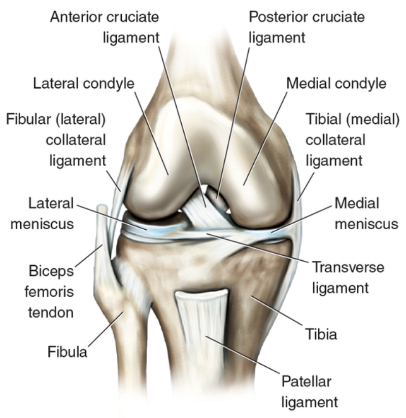
3 bones come together at the knee joint
- Shin bone (Tibia)
- Thigh bone (Femur)
- Kneecap (Patella)
There is also a 4th bone the fibula which is located next to the tibia and the knee joint.
The knee joint is also made up of 4 ligaments which are thick bands of tissue that stabilise the knee joint these are:
- The Medial Collateral Ligament (MCL)
- The Lateral Collateral Ligament (LCL)
- The Anterior Cruciate Ligament (ACL)
- The Posterior Cruciate Ligament (PCL)
The knee joint contains 2 large pieces of cartilage called menisci which prevent the bones from rubbing together. The knee joint has 3 bursae which act as shock absorbers and cushion the knee during movement. These are located above, below and in front of the knee cap (patella).